Perinatal stem cells are obtained around the time of birth, either shortly before or after. They can be sourced from umbilical cord blood and tissue, placental blood and tissue, as well as amniotic fluid and tissue.
When stem cells were first identified, only two types of stem cells were described: embryonic stem cells and adult stem cells. Today, a diverse range of stem cells exist, because the diversity of adult stem cell types has substantially expanded and induced pluripotent stem cells (iPSCs) have since been discovered.
Researchers debate the number of stem cells in existence, because the final figure depends on whether stem cells are characterized functionally (for example, neural stem cells, mesenchymal stem cells, hematopoietic stem cells, etc.) or characterized by source tissue (for example, cord blood stem cells, dental pulp stem cells, amniotic stem cells, etc.).
What are Perinatal Stem Cells?
Perinatal is a term that defines the short period right before to right after birth. Perinatal stem cells encompass both pre-natal and post-natal stem cells.
To classify stem cell types, you can apply the following definitions:
- Adult stem cells – Stem cells derived from living humans
- Embryonic stem cells – Stem cells derived from embryos
- Pre-natal stem cells – Stem cells derived from the fetus or supporting structures
- Post-natal stem cells – Stem cells derived from a recent newborn
Based on those definitions, the primary characteristic that separates embryonic vs. perinatal vs. adult stems cells is their source tissue. However, it is possible to further characterize each stem cell type by another characteristic, which is their differentiation capacity.
For example, stem cells can be further characterized by whether they represent a population of totipotent, pluripotent, or multipotent cells, as described below.
Totipotent vs. Pluripotent vs. Multipotent
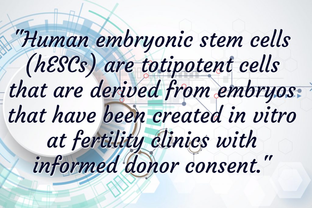
Human embryonic stem cells (hESCs) are totipotent cells that are derived from embryos that have been created in vitro at fertility clinics with informed donor consent. Embryonic stem cells are typically harvested shortly after fertilization (within 4-5 days) by transferring the inner cell mass of the blastocyst into a culture medium. In mammals, the fertilized oocyte, zygote, 2-cell, 4-cell, 8-cell and morula resulting from cleavage of the early embryo are all examples of totipotent cells.[1]
However, at 5-6 days post-fertilization, totipotent cells begin to specialize, at which point they become pluripotent or multipotent cells.
Pluripotent and multipotent stem cells have a more limited differentiation capacity than totipotent stem cells. As an example, multipotent blood stem cells can differentiate into red cells, white cells and platelets in the blood, but they cannot become any cell type.
Stem cell definitions based on differentiation capacity are provided below:
- Totipotent stem cells – Cells that have the capacity to form an entire organism
- Pluripotent stem cells – Can give rise to most, but not all, tissues within an organism
- Multipotent stem cells – Undifferentiated cells that are limited to giving rise to specific populations of cells
The precise point at which a stem cell switches from a totipotent stem cell to a pluripotent or multipotent stem cell can sometimes be unclear. Furthermore, technology now allows us to reverse engineer mature cell types back into a totipotent state. This cell type is called an induced pluripotent stem cell, also known as an iPSC or iPS cell for short.
iPS cells are totipotent, so the scientific community has essentially come full circle in our ability to take cells those these various stages of development.
Perinatal Stem Cells: Pre-Natal and Post-Natal Stem Cells
Perinatal is a term describing the period right before and after birth, so it includes both prenatal and post-natal stem cells. Several types of perinatal stem cells are described below.
1. Gonadal Ridge, 6 months (Pre-natal; Pluripotent)
Primordial germ cells exist briefly in an embryo before they associate with somatic cells of the gonads and differentiate into germ cells. Human embryonic germ cells (hEGCs) are stem cells that originate from the primordial germ cells of the gonadal ridge of a 5- to 9-week old fetus. Since 1998, hEGCs have been successfully isolated and characterized.[2] They are pluripotent because they can develop into any of the three potential germ layers: ectoderm, mesoderm, and endoderm.
2. Fetal Stem Cells (Pre-natal; Pluripotent)
While early hESCs and gonadal ridge hESCs are derived from pre-implantation embryos, fetal stem cells are derived from primitive cells in the organs of fetuses. Specifically, neural crest stem cells, fetal hematopoietic stem cells and pancreatic islet progenitor cells are three types of stem cells that have been isolated from fetuses.[3] Fetal stem cells are characterized by pluripotency, with neural stem cells taken from fetal brain tissue able to differentiate into both neurons and glial cells,[4],[5] and hematopoietic stem cells isolated from fetal blood and placenta able to differentiate into several blood cell types.
3. Cord Blood Stem Cells (Post-Natal; Multipotent)
The blood present in the newborn umbilical cord contains circulating stem cells. There are several important properties of umbilical cord blood, including that umbilical cord blood hematopoietic stem cells equal or exceed the frequency of those in bone marrow, they can produce large colonies in vitro, they have different growth factor requirements, and they can be expanded in long-term culture.[6]
Cord blood stem cells are characterized as multipotent, as they are capable of differentiating into numerous stem cell types, including neurons, hepatic cells, and circulating cell types.[7]
Typically, umbilical cord stem cells are most easily differentiated into cells found within the blood and lymph (immune) systems, such as red blood cells, which transport oxygen within the body; white blood cells, which combat bacterial and viral infection; and platelets, the “sticky” cells that aid in the clotting of blood.
The ability of cord blood stem cells to differentiate into cells of the blood and immune systems means that they hold significant potential for use in the treatment of diseases that include heart disease, stroke, and neurological conditions like Alzheimer’s.
4. Cord Tissue Stem Cells (Post-Natal; Multipotent)
While most research up to this point has focused on the blood present in umbilical cord tissue, the matrix cells that form umbilical cord tissue (known as “Wharton’s Jelly”) also contain stem cells. Wharton’s Jelly has been a source of isolation for mesenchymal stem cells, which express typical stem cell markers (such as c-kit and high telomerase activity). Mesenchymal stem cells derived from this source have been propagated for long population doubling times and can be induced to differentiate in vitro into neurons[8].
To date, this area of stem cell research has not produced FDA approved clinical applications, although several clinical trials are underway. Two particularly promising applications for this type of stem cell (among many other potential applications) include:
- The ability of umbilical cord matrix stem cells to express hepatic markers and differentiate into hepatocyte-like cells[9],[10],[11]
- The ability of umbilical cord matrix stem cells to differentiate into neurons and glia[12],[13] ,[14]
5. Placental Stem Cells (Post-Natal; Multipotent)
As described by placental stem cell expert, Dr. Ornella Parolini, placenta-derived stem cells include several different types of cells, such as hematopoietic stem cells derived from cord blood and all the cells derived from the placental tissue, which include amniotic and chorionic membranes, the chorionic villi, the umbilical cord, and the maternal component which is the decidua.

Advantages of placental stem cells include their early embryological origin, high cell yield, and the fact that placental cells have been less exposed to infections (they come from an infection-naïve environment), which render them less likely to transmit disease.
Summary of Perinatal Stem Cells
Although great controversy has surrounded embryonic stem cells (ESCs), an extensive range of stem cell types can be ethically sourced from living human beings. Nonetheless, the process of sourcing each cell type and their differentiation capacity can vary. For those operating in the stem cell sector, having a clear understanding of the characteristics associated with each stem cell type can pave the wave for new research inquiries, novel diagnostic tools, and innovative therapeutic applications.
To learn more about market for perinatal stem cells, view the “Global Cord Blood & Tissue Banking Industry Report – Market Size, Segmentation, & Forecasts.”
Footnotes:
[1] Bongso A, Richards M. History and perspective of stem cell research. Best Practice & Research Clinical Obstetrics & Gynaecology. 2004; 18(6): 827-842.
[2] Shamblott MJ, Axelman J, Wang S, et al. Derivation of pluripotent stem cells from cultured human primordial germ cells. Proc Natl Acad Sci USA. 1998; 95: 13726–13731.
[3] Beattie GM, Otonkoski T, Lopez AD, et al. Functional beta-cell mass after transplantation of human fetal pancreatic cells: Differentiation or proliferation? Diabetes. 1997; 46: 244–248.
[4] Brustle O, Choudary K, Karram K, et al. Chimeric brains generated by intraventricular transplantation of human brain cells into embryonic rats. Nat Biotech. 1998; 16: 1040–1044.
[5] Villa A, Snyder EY, Vescovi A, et al. Establishment and properties of a growth factor dependent perpetual neural stem cell line from the human CNS. Exp Neuro. 2000; 161: 67–84.
[6] Rogers I, Casper RF. Umbilical cord blood stem cells. In Best Practice &Research Clinical Obstetrics&Gynaecology. 2004; 18(6): 893-908.
[7] Ilancheran, et al. Human fetal membranes: a source of stem cells for tissue regeneration and repair? Placenta. 2009; 30(1): 2-10.
[8] Bongso A, Lee EH. E-textbook: Stem Cells: Their Definition, Classification and Sources. http://www.worldscibooks.com/lifesci/etextbook/5729/5729_chap1.pdf. Retrieved March 20, 2014.
[9] Campard, et al. Native umbilical cord matrix stem cells express hepatic markers and differentiate into hepatocyte-like cells. Gastroenterology. 2008; 134 (3): 833-848.
[10]Sáez-Lara, et al. Transplantation of human CD34+ stem cells from umbilical cord blood to rats with thioacetamide-induced liver cirrhosis. Xenotransplantation. 2006; 13(6): 529-535.
[11] Wang, et al. Induction of umbilical cord blood-derived beta2m-c-Met+ cells into hepatocyte-like cells by coculture with CFSC/HGF cells. Liver Transpl. 2005; 11(6): 635-643.
[12] Mitchell, et al. Matrix Cells from Wharton’s Jelly Form Neurons and Glia. Stem Cells. 2003; 21: 50-60.
[13] McGuckin, et al. Culture of embryonic-like stem cells from human umbilical cord blood and onward differentiation to neural cells in vitro. Nat Protoc. 2008; 3(6): 1046-1055.
[14] Jomura, et al. Potential treatment of cerebral global ischemia with Oct-4+ umbilical cord matrix cells. Stem Cells. 2007; 25(1): 98-106.