This guest post by JM Wilkinson and Kelly S. Davidge of Kirkstall Ltd. highlights an emerging technology, organotypic cell cultures.
Cell culture has come a long way in the last 20 years. 3D is rapidly replacing 2D (plated cells on cover slips) and now perfusion based systems with gentle flow of media are improving proliferation rates, control of differentiation and cell function. For in vitro safety and efficacy testing the goal is organotypic behaviour where the cell metabolism accurately reflects what happens in the human body. However some of the techniques also have direct relevance for stem cell research too. In this note we discuss some of the important factors that help to steer stem cell differentiation and could deliver mature cells with low batch to batch variation and a far more homogeneous population.
Current protocols for differentiating IPSCs usually involve complex sequences of culture with addition of expensive growth factors at critical time points. The techniques of organotypic cell cuture suggest that by controlling the environment of cells more carefully we may be able to increase proliferation rate without risking epigenetic mutations. These are some of the factors that we need to control:
- Oxygen stress
- Flow stress
- Extracellular matrix
- Feeder Cells.
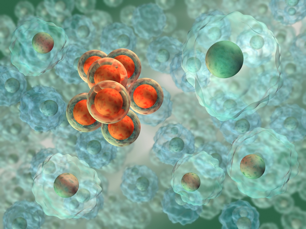
Oxygen levels in cell culture are usually 20% at the surface of the media and falling in the depth of the culture vessel dependent on the metabolism rate of the culture. Culture plates are often rocked or shaken to mix the media and achieve a better supply of oxygen to the cells but at the expense of creating turbulent flow and inducing variable flow stress in the growing cells. This is certainly one cause of divergence in differentiation path and is illustrated clearly by spheroid culture where cells on the periphery become fibroblast-like, whereas away from the periphery the cells maintain their morphology and those in the centre may be in hypoxic conditions and suffer from necrosis.
The latest platform for organotypic cell culture, the Quasi Vivo® system, enables the oxygen concentration, medium flow rates and flow stress to be accurately modelled and precisely controlled. This allows the researcher to know precisely what is happening inside the chambers and allows exact control of variables in an experiment. Importantly, low levels of flow stress itself has been shown to be beneficial to cells as it more closely mimics their natural environment, and allows cells to behave in a more normal fashion.
A second important factor is the provision of an extracellular matrix (ECM) that helps build 3D tissue-like cultures instead of maintain the cells in suspension (which is un-natural for all but a few cells such as those in the blood stream). Organotypic cell culture is moving away from simple rigid scaffolds to compliant gels which not only have the right mechanical properties but can be loaded with the right proteins which act as a synthetic ECM until the cells generate their own matrix.
The Quasi Vivo® system provides a new platform for cell culture that has already demonstrated its value in supporting liver models as well as co-culture of cardiomyocytes, MSCs and models for the gut and blood brain barrier. It is a modular system where the number of chambers in culture can be expanded. The system supports culture of cells in suspension, cells on scaffolds, spheroids, and even thin tissue slices with maintenance of viability and function over 28 days.
In traditional stem cell culture, feeder cells are often required to support the growth of stem cells, which they do by secreting molecules and cytokines into the medium to encourage expansion of iPSCs, and are included in the same vessel as the stem cells. Separating these two cell populations for differentiation or analysis can involve lengthy protocols such as serial passaging, and new techniques are being developed which include the use of FACs or labelling to distinguish between the two cell populations.
The Quasi Vivo® system provides a natural solution to this problem: iPSC and feeder cells can be cultured in separate chambers, linked together via perfusion flow. In this set up, molecules, cytokines and proteins expressed by the feeder cells flow freely through the system, including into the chamber containing the iPSCs, and no separation step is required; one could simply disconnect the feeder cells from the loop, and harvest or continue culturing the iPSCs as required.
About the Authors:
Malcolm is a veteran of the microelectronics industry who, after twenty years of designing microchips, has finally found something more rewarding – designing ‘organs on chips’ to help develop safer medicines without using animal testing. His engineering and systems approach to designing medical and biological products has been a feature of the short courses he has presented on behalf of the Swiss training organisation FSRM over the last 10 years. He now runs a start-up company in Sheffield UK.
Kelly is Research and Development Manager at Kirkstall Ltd. Her BSc was in Microbiology from Cardiff University, with PhD studies at the University of Sheffield. Following on from Post Doctoral research posts at the Universities of Sheffield and Nottingham, she joined Kirkstall in January 2015 and is working with academia and industry to develop alternatives to animal testing through the innovative application of in vitro technology.
About Kirkstall:
Kirkstall is a biotechnology company specializing in products and services supporting in-vitro cell culture research and the development of routine in vitro testing. The company’s mission is to offer more accurate and reliable predictive models of human physiology without the use of animals. Visit http://kirkstall.org/ to learn more.