During the fall months of cancer campaigning, perhaps the biggest awareness remains in shadow: it is not yet possible to prevent patients from receiving needless treatment. As it stands, conservative estimates have determined that greater than 50% of men and women undergo unnecessary surgery and chemotherapy[1]. Many cancers do not deserve the dreaded name and are beginning to be labeled as non-progressive or indolent (“IDLEs”)[2] –a categorization that by itself may prevent untold psychological and physical trauma.
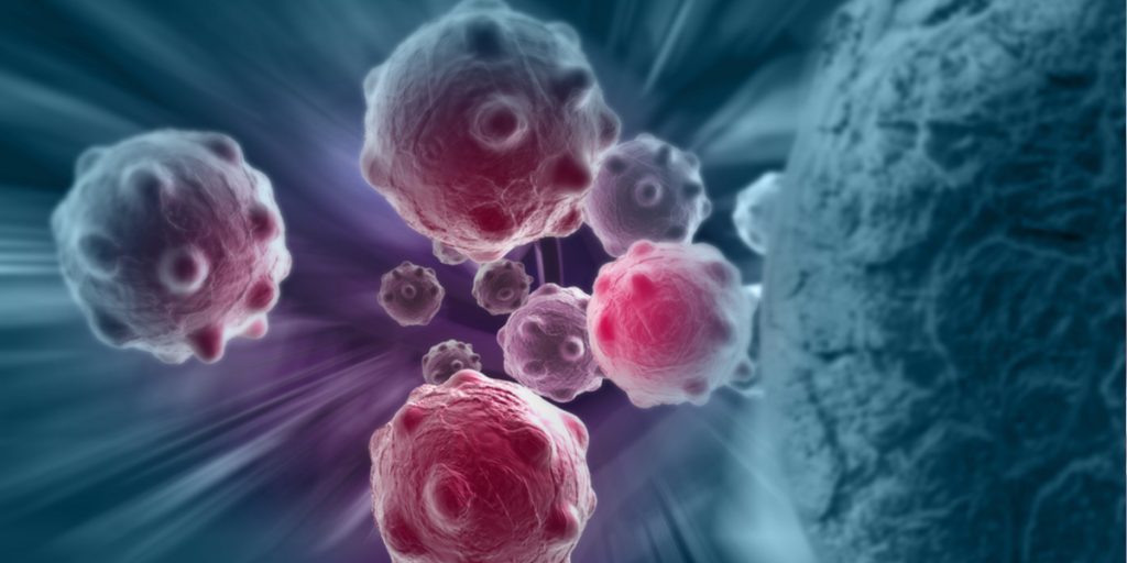
Exosome Diagnostics May Predict Invasiveness
Savvy researchers recognize that the most accurate information providing that distinction will come from what cells release into the blood like molecular mail[3]. Cancer or IDLEs, regardless of threat level, release clues regarding structural rearrangements, as well as tell-tail enzymes that allow cell migration, commandeer local metabolism, and resist their pollution.
Profiling these clues would enable scientists and doctors to determine the necessity and precision of therapy, rather than automatically prescribing aggressive treatment.
Such mail is not released “naked” but in little parcels of cell membrane called extracellular vesicles (EVs) or exosomes. Exosomes are produced by all cells in day to day bodily functions, but on a cell-for-cell basis, cancers release larger quantities of exosomes[4].
Using Exosomes to Identify Cancer Severity
In a study recently published by Professor Alan Doucette (Dalhousie University Nova Scotia) with Dr. Aled Clayton (Cardiff Medical School U.K.) Michelle Cormier (ACRI Moncton; now RCMP Ottawa) and Dr. Steve Griffiths (Minervagen) a simple method can sort the post and open it more quickly. 22 years ago, Dr. Griffiths while at Moncton’s Atlantic Cancer Research Institute (ACRI), learned from pioneering exosome expert Dr. Clayton that exosomes released by cancer cells are tied up with chaperone proteins.
The chaperones are a universal requirement of cancers regardless of threat level to maintain the hyperactivity of distorted and displaced proteins; these allow them to ignore normal rules, consume and utilize more nutrients and survive a self-polluted environment.
Dr. Griffiths designed small stretches of protein building-block fragments called peptides with a double headed feature enabling them to stick to different types of chaperones. He called them Venceremins (Vn) from the Spanish expression “Venceremos!” meaning, “We will win.” The size, shape, and number of chaperone ribbons decorating cancer exosomes can vary substantially. However, all of them get cross linked by the peptide.
The chaperone ribbons are not present on the surface of the exosomes produced by normal cells [5]. Once the intercepted parcels are opened, it is possible to examine the instructions and tools inside and whether the cells that produced them need immediate attention or if monitoring would be sufficient [6].
Dr. Clayton was the first to fully appreciate the power of Dr. Griffith’s approach, producing irrefutable evidence for removal of exosomes from clinical samples (something that continues to be impossible even with the most sophisticated and time-consuming approaches). Drs. Clayton and Griffiths met and discussed their exciting results at the first International Exosome Conference at the Marie Curie Institute in Paris 2011. “I couldn’t have been happier, “said Dr. Clayton.
Crucially, separation of exosomes can be carried out easily and quickly in a short spin by a centrifuge. The exosomes are collected as a thin sediment at the bottom of a test tube. This leaves most of the unwanted material suspended in fluid and easy to remove.
As a bonus, the exosomes are so tightly tethered that they can be resuspended and further washed to remove non-specific material with detergents. Now purged, the exosomes can be opened and analysed for evidence of malignancy.
Using Exosome Proteins to Identify Invasive Cancer Cells
In the study, exosomes were captured from bioreactor cultures that provide more natural conditions than traditional flasks or plates normally used for growing cells. In bioreactors, cells pile on top of each other and grow in layers and islands; they may be maintained for over 4 months. Traditional cultures consist of single layers of cells and are discarded after a few days after they have reached the edge of their dish (confluence). The authors regard the distinction with some importance in trying to determine what types of exosomes might be produced in “real life”.
For comparative purposes, captured exosomes were captured from the culture medium of bioreactors containing a normal “fibroblast” or skin like culture (benign) and compared to a non-invasive breast cancer culture (ductal carcinoma in situ; DCIS) and an invasive culture that mimics breast cancer when injected into laboratory animals.
Professor Doucette of Dalhousie University is an internationally renowned expert in proteomics. He used a device called a mass spectrometer to profile total protein content to at least distinguish between the normal and cancer cells. Surprisingly, it became quickly apparent that exosomes from the invasive breast cancer could be distinguished not only from those of normal cells, but also from the non-invasive source.
One after another, the most abundant exosome proteins released by the invasive cells could be confirmed as biomarkers identified in independent studies, many of which had only been confirmed in the past year. The trees leapt out of the forest. It was a snapshot of important clues that could be captured all at the same time.
The Future of Cancer Treatment
The Vn peptides will bind to all types of cancer exosomes. The approach may bring new appreciation for common exosome contents produced by malignant cells and how to treat them. Cancer may be further exposed as a vulnerable entity and not infinitely variable. It is the identification of common threads in the varied tapestries rather than a magic bullet.
“If there’s one message I would like to give to cancer researchers” said Dr. Griffiths, “If you have any cell cultures in your lab, find the time to take a few days, sample some cell culture medium–you don’t have to sacrifice the culture even–and send it off for protein analysis. You will be amazed at what they are telling you about how they work and what you might look for in the patient.”
In the interim, some labs around the world are already using the approach on clinical samples, including a group in the Netherlands headed by Prof. Dr. Connie R. Jimenez Head OncoProteomics Laboratory, VUmc-Cancer Center Amsterdam. Professor Jimenez and her teams are looking at detection of exosomes released into the urine, as well as into the blood of patients with prostate and colorectal cancer. Dr. Jimenez shares, “Steve’s peptides are catalyzing all our exosome proteomics projects in a variety of cancers.”
The paper can be downloaded free of charge at the following link.
Footnotes
[1] Effect of Three Decades of Screening Mammography on Breast-Cancer Incidence Bleyer, M.D (2012) Engl J Med 2012; 367:1998-2005
Paradigm Shift toward Reducing Overtreatment of Ductal Carcinoma In Situ of Breast. Sagara YFront Oncol. 2017 Aug 28;7:192.
Benefits and harms of breast cancer screening with mammography in women aged 40-49 years: A systematic review. van den Ende Int J Cancer. 2017 Oct 1;141(7):1295-1306
[2] “We propose the term indolent lesion of epithelial origin, or IDLE, for those lesions (currently labelled as cancers) and their precursors that are unlikely to cause harm if they are left untreated.” Addressing overdiagnosis and overtreatment in cancer: a prescription for change. (2014 ) Esserman LJ, Lancet Oncol. May;15(6):e234-42
[3] “The Secretome”: Integration of Breast Cancer Secretomes with Clinical Data Elucidates Potential Serum Markers for Disease Detection, Diagnosis, and Prognosis. 2016 Ziegler YS PLoS One. Jun 29;11(6):e0158296.
[4] These can fuse to normal neighbor cells and make them work differently to their own desired metabolism; some circulate through blood to “seed” for secondary growth known as metastasis.
[5] Even “normal” cells– rapidly growing in the laboratory– have a different set of chaperones because and these can be collected too for comparison to the different stages of cancer cells (although they release about 25% less mail).
[6] Why doctors are rethinking breast cancer treatment (TIME)
Ready to learn more about this exciting market area? View the “The Global Exosome Market – Market Size, Forecast, Trials and Trends.”