Neurodegenerative diseases, like Alzheimer’s and Parkinson’s disease are caused by progressive damage to cells of the nervous system. Patients suffering from these debilitating disorders lose their mobility, balance, speech, and so many other important functions. Scientists studying these disorders need advanced cell models that have the physiological and pharmacological properties seen in mature neurons.
Human induced pluripotent stem cells (iPSCs) with their extensive differentiation potential are valuable tools for understanding the pathology of neurodegenerative disorders. Neuronal cells that are derived from iPSCs have the required physiological properties seen in mature neurons from the human brain. Rodent-derived models can’t quite do that, and human tissue is mainly available post-mortem. Researchers can use iPSCs to explore neurite length and cell health, drug induced effects on morphology, neuronal activity, among other questions.
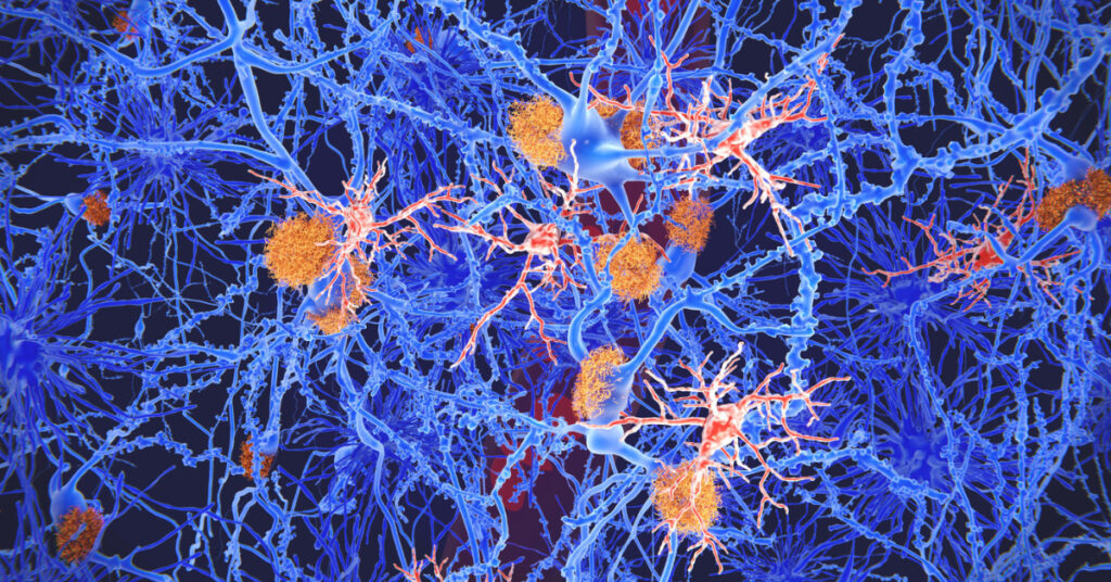
Cells of the immune system, like glial cells, also regulate neuronal networks and can play a role in the development neurodegenerative disorders. Microglia, for example, are the primary immune cells of the nervous system. These macrophage-like cells are the front line of defence against injury or infection in the central nervous system. Neuroinflammation is the distress signal that activates microglia, which then phagocytose unhealthy neurons. Prolonged activation of microglia, however, is harmful to neuronal networks and is linked to neurodegeneration.
Axol Biosciences is a supplier of validated human iPSC-derived cells and primary cells for research on neurodegeneration and inflammation. We spoke with Dr. Sian Humphries of Axol Biosciences about their work and the vital role of live-cell imaging and analysis in their field.
Interview with Dr. Sian Humphries
Sartorius: Could you describe the research models commonly used in your company, as well as the workflows that are important in the field of iPSC cell line development from an industrial perspective?
Dr. Humphries: We have expertise in differentiation of iPSC to cortical, motor and sensory neurons, macrophage, microglia and astrocytes. We run assays relevant to neurodegeneration and inflammation and are always developing new models, such as co-cultures. We have access to a range of iPSCs and tissue for reprogramming, carrying disease-relevant mutations.
Glia cultures of astrocytes, microglia, and oligodendrocytes developed using primary cultures or iPSCs are an important research tool in the field. Studies using these models benefit from advanced analytical tools for understanding glia cell growth, morphology, function, and ultimately their roles in diseases of the central nervous system.
Sartorius: Let’s focus on glia, particularly on microglia, and its role in neuroimmune diseases. Could you describe what phenotypes of interest one may look for when developing human iPSC- derived microglia, and what research applications are best suited to enable their characterization, from morphology to functional assessment?
Dr. Humphries: We have worked with microglia derived from TREM2 and CD33 variant iPSCs, which are obviously relevant to neurodegeneration. In particular, phagocytic and chemotactic responses are of interest, and we also measure release of cytokines.
Sartorius: Moving onto another very important glia cell, astroglia, and their interaction with neurons, how do you think glia cells shape neuronal activity? Also, what is the best way to model this interaction in a research environment?
Dr. Humphries: Astrocytes play a key protective role for neurons. As we are able to use Incucyte® Neurolight and Incucyte® Neuroburst reagents to distinguish between astrocytes and neuronal cells, assays such as the neurite outgrowth and spontaneous neuronal activity can be run in the presence of astrocytes and other cell types.
Human iPSC-derived cell models are widely used in basic neuroscience research, drug development, and neurotoxicity studies. Traditional endpoint methods are limited in answering fundamental questions about how these cells differentiate, mature and become functional. Having to transfer culture plates from the incubator to analytical instruments can also be harmful to cells and their function.
Sartorius: To wrap up, can you spare a few thoughts on the future of drug development and screening assays?
Dr. Humphries: Continuous imaging facilitates optimization of incubation time and reduces the number of plates of cells used. The ability to perform multiple assays simultaneously adds further depth to the data. We anticipate more complex co-culture models and multiple readouts, such as cell health in tandem with other functional readouts.
Conclusion
The ability to monitor the health, morphology and behavior of cells of the nervous system in real time, without having to remove them from the incubator is paramount to the research and development of effective treatments for neurodegenerative diseases. The Incucyte® Live-Cell Analysis Systems, with Neuroscience analysis software are compatible with advanced cells models, like iPSC-derived or primary cells, in either mono- or co-culture, bringing together a powerful combination of more physiologically relevant disease models and real-time live-cell analysis.
The next generation of therapies for neurodegenerative disorders awaits…